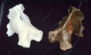
This 3D model was rendered from a CT scan. The original specimen was recovered by W.D. Frankforter in 1959 near Dunlap, IA and is in the collection of the Sanford Museum and Planetarium, Cherokee, IA.
Note: It is January 2014 and this blog has been sitting stagnant for quite awhile. Over the next few months, I have a number of posts planned. Most are about the paleontology/paleoecology of Ice Age mammals, but I’m planning a few that will focus on methods and documentation–especially for the amateur community. So stay tuned!
A few weeks ago, this article appeared in the Springfield Journal Register. I thought it was a very good article–evidently my head even made it “above the fold” on the front page (which I’m told is a good thing). I’m always surprised at the “big deal” factor of 3D. Yes, some of it may be because the technology is coming down in price, and the free and open source (FOSS) software resources are out there–and very good. But part of me hopes that it is something bigger, a profound change in the way we think about museum objects and who has access to them.
For my part, thinking in 3D is simply an extension of what we’ve always done. Bone morphology (size and shape) is the bread and butter of most paleontologists. We are always trying to tease out the reasons why a bone is this species rather than that species, and 99% of the time, the answer hinges on its shape. The new 3D technologies are making this easier. So we’ve been scanning specimens from our collections–almost constantly–for the last few months. Everything from Dire Wolves, to Tully Monsters, Mastodon feet to trilobites. At this stage, we’re still in the “let’s see how this works” phase, but soon, perhaps very soon, we’re hoping to put many of these 3D models on the web. We’re in good company here. The Smithsonian released a number of 3D scans that can be found here, including a very nice woolly mammoth (it is 3D printer ready!). We’re almost there.
But why would a person want to 3D print a mastodon foot? How might scanned museum specimens be useful to, well, everybody else? Museums have been in the replicating business for a long time. We mold and cast our most significant specimens so they can be shared with researchers and other museums. However, this “analog” way to replicate fossils was sometimes imprecise, and definitely required quite a bit of skilled time and labor. For a number of years, we made mastodons and ground sloths to order. They weren’t cheap, and they were never a money-making proposition. But they provided a valuable service to museums that were not as rich in collections as we are.
Fast forward to the 21st century. Awhile back we reported a very interesting saber-toothed cat find in southeastern Minnesota Cave (see a great blog post on it here). The Minnesota Karst Conservancy, who owns the cave (and therefore the fossils) preferred that the fossils stay in the state. So after our analysis is complete, they will be returned to the Science Museum of Minnesota where an exhibit awaits. Because we spent a lot of time analyzing these materials, I wanted to retain casts here at the Illinois State Museum. In the future, when researchers visit to look at other similar specimens, they can at least capture basic measurements on fossils from this locality. When it came time to discussing making casts of the major bones, it soon became clear that a traditional mold/cast would sacrifice detail and be very labor intensive. Being the sort of person who likes to avoid hard work for the sake of work, I started dreaming up alternatives. Ultimately, we scanned most of the major bones in a CT scanner at a local hospital. The resulting images could be rendered as a 3D model and printed on a 3D printer at MAD Systems in CA (here).
But that’s not the only reason to 3D print things. A physical object can be manipulated and examined in ways that photos cannot. Our current scanning efforts have focused on the most significant fossils in our collections, including: some of North America’s earliest dogs (Koster Site, Greene Co., IL) and the continent’s largest Mastodont (from Boney Spring, MO). It is our job to see that the public benefit and learn from our collections. That they don’t sit on a shelf, forgotten. The more people that can enjoy these objects for themselves, the better!
Where are we going with this technology? We’re not entirely sure yet. Certainly, we will integrate this into ongoing efforts to digitize our collections. We will work 3D models into our outreach and education activities. Will there be more? Undoubtedly. Stay tuned to our FB page and the Think3D website for new developments!
